Abdominal ultrasound ​

What is a private abdominal ultrasound?

Procedures

Ultrasound

What is an abdominal ultrasound?

A private abdominal ultrasound is an ultrasound examination of the abdomen that is performed by a private clinic or hospital, rather than a publicly funded healthcare facility. Patients typically choose a private abdominal ultrasound for a number of reasons, including:

  • Quicker access to an appointment: Private healthcare facilities can often provide faster access to an ultrasound appointment compared to the public healthcare system, as there may be shorter waiting lists.
  • Increased comfort and privacy: Private clinics and hospitals often offer more comfortable and private facilities compared to the public healthcare system.
  • Increased control over the examination: Patients can choose the type of private abdominal ultrasound they would like to have, and they may be able to request a specific radiologist or sonographer.
  • More detailed reports: Private clinics and hospitals may offer more detailed reports and results compared to the public healthcare system.
  • Increased flexibility: Patients can often choose the time and date of their private abdominal ultrasound appointment, which can be particularly important for those who have busy schedules.

However, it is important to note that private abdominal ultrasounds are typically not covered by healthcare insurance and must be paid for out-of-pocket by the patient. Additionally, private abdominal ultrasounds may not always be necessary, and a referral from a GP or specialist may be required.

What can you see during an abdominal ultrasound?

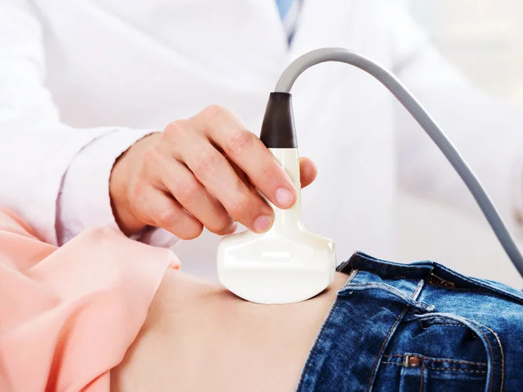
An abdominal ultrasound is a medical imaging test that uses high-frequency sound waves to create images of the organs and tissues within the abdomen. During an abdominal ultrasound, a doctor or a  sonographer (technitian) applies a special gel to the patient’s skin and uses a handheld probe to generate the sound waves and capture the images.

An abdominal ultrasound can provide valuable information about the following organs and structures:

  • Liver: The ultrasound can assess the size, shape, and texture of the liver and detect any abnormalities, such as cysts, tumors, or cirrhosis.
  • Gallbladder: The ultrasound can detect gallstones, evaluate the function of the gallbladder, and identify any inflammation or infection.
  • Pancreas: The ultrasound can evaluate the size and shape of the pancreas and detect any abnormalities, such as cysts or tumors.
  • Spleen: The ultrasound can assess the size and texture of the spleen and detect any abnormalities, such as an enlarged spleen or a mass.
  • Kidneys: The ultrasound can evaluate the size, shape, and texture of the kidneys and detect any abnormalities, such as cysts, tumors, or kidney stones.
  • Abdominal blood vessels: The ultrasound can evaluate the blood flow in the major arteries and veins within the abdomen, such as the aorta and the vena cava.
  • Abdominal fluid collections: The ultrasound can detect the presence of fluid within the abdomen, such as ascites, which can be a sign of liver disease or other underlying conditions.

In addition to these organs, an abdominal ultrasound can also help diagnose other conditions such as hernias, abdominal pain, and digestive tract problems. The images obtained during an abdominal ultrasound can help guide further diagnostic tests or treatments as needed.

What is a pelvic ultrasound ?

A pelvic ultrasound is a non-invasive diagnostic imaging test that uses high-frequency sound waves to produce images of the structures and organs within the pelvic area, such as the uterus, ovaries, bladder, and prostate gland.

In gastroenterology, a pelvic ultrasound may be used to evaluate the lower gastrointestinal tract, including the rectum and anus, and to assess the structures adjacent to it, such as the bladder and prostate in males, and the uterus and ovaries in females.

Some of the common uses of pelvic ultrasound in gastroenterology include:

  1. Evaluating the extent of rectal cancer: Pelvic ultrasound can help determine the size and location of rectal tumors, and can also identify any nearby lymph nodes that may be affected.

  2. Diagnosing pelvic floor disorders: Pelvic ultrasound can help diagnose pelvic floor disorders, such as fecal incontinence or rectal prolapse, by assessing the anatomy and function of the pelvic muscles.

  3. Assessing prostate enlargement: In males, pelvic ultrasound can be used to assess the size of the prostate gland and to identify any abnormalities, such as enlargement due to benign prostatic hyperplasia or the presence of tumors.

  4. Evaluating urinary incontinence: Pelvic ultrasound can be used to evaluate the bladder and urinary tract to determine the cause of urinary incontinence.

Overall, pelvic ultrasound is a useful diagnostic tool in gastroenterology, as it provides detailed images of the pelvic organs and structures, allowing physicians to accurately diagnose and treat a wide range of gastrointestinal conditions.

Schedule an appointment

What is a private abdominal ultrasound? Read More »

Transnasal Gastroscopy

What is transnasal gastroscopy?

Procedures

What is transnasal gastroscopy?

Transnasal gastroscopy (TNG) is a minimally invasive endoscopic procedure used to examine the inside of the upper gastrointestinal (GI) tract, including the esophagus, stomach, and duodenum. Unlike traditional gastroscopy, which is performed through the mouth and requires sedation, TNG is performed through the nose and does not require sedation. During the procedure, a flexible endoscope with a tiny camera on the end is passed through the nasal passages and into the GI tract, allowing the physician to visually inspect the inside of the upper GI tract. TNG is often used to diagnose and treat conditions such as acid reflux, ulcers, and other gastrointestinal problems.

Schedule an appointment

What is transnasal gastroscopy? Read More »

colonoscopy

What is a private colonoscopy ?

Procedures

What is a private colonoscopy ?

A private colonoscopy is a medical procedure in which a patient pays for a colonoscopy through a private health insurance policy or out-of-pocket, rather than through the National Health Service (NHS) in the United Kingdom.

A colonoscopy is a procedure in which a thin, flexible tube with a camera at one end (called a colonoscope) is passed into the rectum and colon to examine the inside of the large intestine. The procedure is used to diagnose and treat conditions such as colon polyps, diverticulitis, and colorectal cancer.

In a private colonoscopy, the procedure is performed in a private hospital or clinic, and the cost of the procedure is usually covered by a private health insurance policy or paid for out-of-pocket by the patient. Private colonoscopies are usually performed by a consultant gastroenterologist, and the patient typically has a shorter waiting time for the procedure than they would if they were on the NHS.

While a private colonoscopy is a more convenient and potentially quicker option, it is also usually more expensive than an NHS colonoscopy. It’s important to carefully consider the cost and benefits of a private colonoscopy and to discuss any questions or concerns with a medical professional before making a decision.

Schedule an appointment

What is a private colonoscopy ? Read More »

Gastroscopy

What is a private gastroscopy?

Procedures

What is a private gastroscopy?

A private gastroscopy is a medical procedure in which a patient pays for a gastroscopy through a private health insurance policy or out-of-pocket, rather than through the National Health Service (NHS) in the United Kingdom.

A gastroscopy is a procedure in which a thin, flexible tube with a camera at one end (called an endoscope) is passed down the throat and into the stomach to examine the digestive tract. The procedure is commonly used to diagnose and treat conditions such as acid reflux, indigestion, and stomach ulcers.

In a private gastroscopy, the procedure is performed in a private hospital or clinic, and the cost of the procedure is usually covered by a private health insurance policy or paid for out-of-pocket by the patient. Private gastroscopies are usually performed by a consultant gastroenterologist, and the patient typically has a shorter waiting time for the procedure than they would if they were on the NHS.

While a private gastroscopy is a more convenient and potentially quicker option, it is also usually more expensive than an NHS gastroscopy. It’s important to carefully consider the cost and benefits of a private gastroscopy and to discuss any questions or concerns with a medical professional before making a decision.

Schedule an appointment

What is a private gastroscopy? Read More »

Oesophageal strictures (narrowing of the oesophagus)

Oesophageal strictures

Conditions

Oesophageal strictures (narrowing of the oesophagus)

Medical conditions, such as chronic acid reflux, can result in other health issues. This is due to the damage inflicted on the lining of the oesophagus by stomach acid over time, as well as the rise of stomach acid and bile up the food pipe.

Medical conditions such as chronic acid reflux, where stomach acid and bile frequently rise up into the food pipe (oesophagus), can lead to other health issues. Over time, stomach acid can damage the inner lining of the oesophagus, causing inflammation that may lead to scarring.

Repeated inflammation of the tissue can lead to a continuous process of healing followed by chronic inflammation and re-healing, ultimately resulting in scarring. If scar tissue accumulates, the oesophagus may develop narrow areas known as strictures. These strictures cause dysphagia, making it difficult to swallow food and, in severe cases, liquids, which can lead to dehydration and weight loss.

Oesophageal strictures are likely to occur in around 10% of patients with gastro-oesophageal reflux disease (GORD), where the symptoms of narrowness in the oesophagus are combined with symptoms of GORD such as heartburn, vomiting, sore throat or a cough. Strictures may also occur in the oesophagus due to viral or bacterial infections, certain medications, the ingestion of corrosive substances or as a result of diseases such as eosinophilic oesophagitis (EoE) and cancer.

Diagnosis of oesophageal strictures may involve swallowing barium while an x-ray is taken of the chest to observe any strictures, or an endoscopy examination where a narrow tube (endoscope) containing a light and a camera is inserted into the oesophagus so that doctors can see inside the food pipe.

The most common treatment for oesophageal strictures is endoscopic dilation, where an endoscope is used to move a tiny balloon or another dilation device into the oesophagus to gently stretch it. Proton pump inhibitors (PPIs) may also be prescribed. However, strictures can still return after a year or so, even with treatment, and multiple dilation sessions may be necessary. In severe cases, surgery may be recommended.

Schedule an appointment

Oesophageal strictures Read More »

Gastritis

Gastritis

Conditions

Gastritis

Gastritis is a gastrointestinal condition characterized by inflammation (redness) of the stomach lining. The condition can arise from various factors, including bacterial infection, which may be asymptomatic in some instances. In others, gastritis may present as feelings of fullness after eating, nausea or vomiting, indigestion, and a burning sensation in the stomach.

On rare occasions, gastritis may result in severe abdominal pain, gastric bleeding (as evidenced by vomiting blood or having black stools), as well as ulcers, growths or tumours in the stomach. Such symptoms arise after regions of the stomach lining have eroded, causing tissue damage from stomach acid.

Gastritis is a digestive disorder characterized by inflammation of the stomach lining, which can result from various causes. For instance, bacterial infection, often asymptomatic, can trigger gastritis. Other symptoms of gastritis may include a feeling of fullness after eating, indigestion, nausea or vomiting, and a burning sensation in the stomach.

In rare cases, gastritis can lead to severe abdominal pain, gastric bleeding (indicated by vomiting up blood or black faeces), as well as ulcers, growths, or tumours in the stomach, caused by tissue damage from stomach acid after areas of the stomach lining have worn away.

Sometimes, chronic gastritis can develop as a result of the immune system attacking the stomach lining. Chronic gastritis can also stem from bacterial infection from Helicobacter pylori (H. pylori), smoking, excessive alcohol consumption, or prolonged use of aspirin or ibuprofen (NSAID painkillers), or from stress, major surgery, a serious illness, or injury.

If you experience indigestion that lasts for over a week, severe pain, or symptoms of gastric bleeding, you should seek medical advice. A doctor may test for chronic gastritis using a stool sample to detect blood in your faeces or the presence of H. pylori bacteria, a breath test to identify H. pylori, a barium swallow, or an endoscope to see inside your oesophagus and stomach.

Lifestyle changes such as avoiding NSAID painkillers and alcohol, not smoking, practicing relaxation techniques, eating smaller meals frequently, and cutting down on irritants such as fried, spicy, or acidic foods can help ease the symptoms of chronic gastritis. Acid-reducing medications can also be beneficial.

Tests

Treatments / Symptom Management

Schedule an appointment

Gastritis Read More »

Diverticular disease and diverticulitis

Diverticular disease and diverticulitis

Conditions

Diverticular disease and diverticulitis

Diverticular disease is a condition in which small pockets or diverticula form in the lining of the large intestine and protrude through the bowel wall. This can result in abdominal pain, which is more pronounced in the lower left side, worsens after eating, but eases later. Other symptoms may include changes in bowel movements such as constipation or diarrhoea and the presence of black stools due to blood in faeces.

If an individual has inflamed or infected diverticula, the medical condition is called diverticulitis, which can lead to severe complications. These complications may include a persistent and intense abdominal pain, a fever of 38C or higher, blood or mucus in faeces, or bleeding from the rectum.

Diverticula, which are small pockets that protrude through the bowel wall, may be present in the large intestine without any symptoms. This condition is known as diverticulosis and is typically discovered incidentally during medical scans for other reasons. The likelihood of developing diverticula and diverticulosis increases with age, affecting roughly 10% of people over the age of 45 and up to 80% of those over the age of 85. About 20% of people with diverticulosis will develop diverticular disease.

Not consuming enough fibre is the primary cause of diverticular disease and diverticulitis. Additional risk factors include smoking, being overweight or obese, consuming excessive amounts of alcohol or red meat, and regular use of ibuprofen or aspirin.

A doctor will conduct a physical examination and blood tests to rule out other conditions with similar symptoms, such as bowel cancer, coeliac disease or irritable bowel syndrome. A CT scan or colonoscopy can diagnose diverticular disease.

Treatment for diverticular disease typically involves increasing fibre intake through foods such as fruits, vegetables, wholegrain cereals, potatoes, nuts and beans. Pain relief with paracetamol is recommended. For diverticulitis, a doctor may suggest a fluid-only diet for a few days followed by a low-fibre diet to allow the digestive system to rest and, once recovered, eating a high-fibre diet. In more severe cases, antibiotics, intravenous fluids for dehydration and surgery may be required.

Tests

Treatments / Symptom Management

Schedule an appointment

Diverticular disease and diverticulitis Read More »

Burning Mouth Syndrome

Burning Mouth Syndrome

Conditions

Burning Mouth Syndrome

This condition is known as burning mouth syndrome, which can cause a persistent, painful burning sensation in the lips, tongue, or other areas of the mouth. It may also be accompanied by a dry or numb mouth, a bitter taste, or changes in saliva production. The symptoms may be constant or intermittent.

Symptoms of burning mouth syndrome

The symptoms of this condition may occur daily, with mild sensations upon waking that worsen throughout the day, or with severe sensations that persist throughout the day from the moment of waking. For some individuals, symptoms may come and go. Eating or drinking may temporarily alleviate the sensations experienced. The symptoms can persist for months or even years without treatment and typically do not result in any physical changes to the appearance of the tongue or mouth.

What causes burning mouth syndrome?

Burning mouth syndrome is a relatively uncommon condition, affecting around 0.75 to 15% of the population according to research. It appears to be more prevalent in women, especially those who are menopausal or aged over 50. The exact cause of the syndrome is not fully understood, but it may result from a dysfunction in the nerves responsible for transmitting sensations from the tongue to the brain.

Why see a burning mouth syndrome specialist?

Long-term symptoms of burning mouth syndrome can lead to other negative effects on a person’s health and wellbeing, such as disrupted sleep patterns, fatigue, frustration, and low mood. For some individuals, it can also interfere with daily activities, such as conversing or enjoying meals with others. Seeking the assistance of a specialist is advisable if you are experiencing burning mouth syndrome. A gastroenterologist specialising in this condition can diagnose and provide appropriate treatment to alleviate your symptoms, allowing you to better manage your condition.

Schedule an appointment

Burning Mouth Syndrome Read More »

Oesophageal cancer and pre-cancerous lesions

Oesophageal cancer and pre-cancerous lesions

Conditions

Oesophageal cancer and pre-cancerous lesions

Oesophageal cancer refers to a malignant growth that affects the oesophagus, also referred to as the food pipe or gullet. The symptoms of this condition can be caused by several digestive health conditions, with varying degrees of severity. Although not all symptoms indicate cancer, it is recommended to seek medical attention for any concerning symptoms.

The oesophagus is a tubular organ made of muscles that connects the throat to the stomach, facilitating the passage of food and liquids. Individuals with oesophageal cancer exhibit abnormal cell growth in their oesophagus, which spreads uncontrollably. This type of cancer can develop at any point along the oesophagus.

Symptoms

In the initial stages, oesophageal cancer may not exhibit any noticeable symptoms. However, as the condition progresses, symptoms may start to appear. The cancer usually progresses slowly and forms a tumour that can obstruct the throat as the cells in the oesophagus multiply.

The primary symptoms of oesophageal cancer are:

In the early stages of oesophageal cancer, there may be no noticeable symptoms. However, as the condition progresses, abnormal cell growth in the oesophagus can create a tumour that could potentially obstruct the throat. Some common symptoms of this cancer include experiencing difficulty in swallowing, feeling as if food is stuck in the throat, persistent clearing of the throat, and oesophageal bleeding which can cause coughing up or vomiting of blood, though this is not common. Additionally, stools may appear darker than usual or bloody. Other symptoms may also be present.

Other symptoms include: 

There are several conditions that can cause the symptoms mentioned above, such as gastro-oesophageal reflux disease (GORD). However, it’s worth noting that having GORD can also raise your risk of developing oesophageal cancer. Therefore, if you experience these symptoms frequently or if your current treatment doesn’t seem to be working, it’s advisable to seek medical attention from your doctor.

Diagnosis

Abnormal cell growth in the oesophagus can result in pre-cancerous lesions, known as dysplasia. During an upper endoscopy, we can extract a sample of this tissue (biopsy) for testing. This test is painless as we numb your throat, and sedation is also an option. To learn more about endoscopy, you can watch this video: “Endoscopy: All You Need to Know”.

If a pathologist identifies signs of high-grade dysplasia, the risk of cancer is higher. Cancer can arise from either of the two main cell types found in the oesophageal lining. As it progresses, cancer may affect the surrounding tissues and organs or spread to other parts of the body. If we detect cancerous cells, we will perform additional tests to determine the stage, grade, and type.

Other tests include a CT scan, PET-CT, endoscopic ultrasound, barium swallow, and laparoscopy. Blood tests may also be necessary. If you experience concerning symptoms, it is important to see your doctor promptly for evaluation.

Types

In medical terms, the type of cell in which cancer originated is known as histology. The two primary histological types of oesophageal cancer are adenocarcinoma and squamous cell carcinoma. Adenocarcinoma is frequently associated with Barrett’s oesophagus and is more common in the UK. In contrast, squamous cell cancer occurs less frequently. Other, less common histological types include small-cell carcinoma, sarcoma, melanoma, lymphoma, and choriocarcinoma. In some cases, we may not be able to identify the specific histological type of cancer, which we refer to as undifferentiated.

Causes

The precise cause of oesophageal cancer remains uncertain. However, it may result from DNA damage in the cells that line the interior of the oesophagus. DNA damage can arise from various lifestyle choices, such as smoking, or prolonged irritation caused by reflux and other medical conditions. Such DNA mutations or alterations may cause cancer. In oesophageal cancer cells, numerous genes may display changes.

Risk factors

The majority of individuals diagnosed with oesophageal cancer are over the age of 60, with men being more frequently affected than women. Other risk factors include being overweight, smoking, or consuming alcohol, particularly more than 14 units per week. Pre-existing conditions such as GORD or Barrett’s oesophagus also increase the risk of developing oesophageal cancer. Additionally, the risk slightly increases after radiotherapy for certain cancers, such as breast, lung, oropharyngeal, or laryngeal cancer. Furthermore, there are reports suggesting that consuming freshly boiled or equally hot drinks may raise the risk of developing oesophageal cancer.

Causes

The precise cause of oesophageal cancer remains unknown. Nevertheless, it is thought to result from DNA damage in the cells that line the interior of the oesophagus. This damage can be caused by certain lifestyle choices, such as smoking, or by chronic irritation from conditions such as reflux. The DNA mutations or changes that result from such damage can lead to the development of cancerous cells. In oesophageal cancer, many genes may undergo changes or mutations.

Treatment

Early stages of oesophageal cancer or pre-cancer can be treated with endoscopic procedures, such as endoscopic resection and HALO radiofrequency ablation, which can remove abnormal tissue and prevent the need for an oesophagectomy (surgical removal of part of the oesophagus). Other treatment options include chemotherapy, radiotherapy, targeted therapy, and immunotherapy.

A multidisciplinary team of highly qualified specialists will work with you to develop a personalised treatment plan based on the stage, grade, and type of cancer.

Schedule an appointment

Oesophageal cancer and pre-cancerous lesions Read More »

High-resolution manometry of the oesophagus

Eosinophilic oesophagitis (EoE)

Conditions

Eosinophilic oesophagitis (EoE)

Eosinophilic oesophagitis, commonly referred to as oesophageal asthma, is an uncommon condition that affects approximately one in 3,000 individuals in the United Kingdom. This disorder results in persistent inflammation of the tissue in the oesophagus, also known as the food pipe. While some individuals experience severe and distressing symptoms, others may only experience mild discomfort.

The symptoms of eosinophilic oesophagitis can manifest differently based on an individual’s age and personal experience. In the context of this page, we will focus on EoE as it occurs in adults. However, it’s worth noting that infants and children are also susceptible to developing EoE.

Related Symptoms

Certain individuals may adapt their eating habits to manage the symptoms associated with EoE. This may include chewing food thoroughly, taking longer than usual to consume a meal, preferring softer foods, or drinking frequently while eating. Additionally, the condition may hinder an individual’s ability to participate in social activities involving food consumption, such as sharing a meal with others.

When to seek emergency help

In the event that food becomes lodged in the oesophagus, it may result in severe circumstances whereby the oesophagus is entirely obstructed. Should you encounter challenges with breathing, experience noisy breathing or choking, it is imperative that you seek emergency assistance.

We recommend that you schedule a consultation with our specialist if you are persistently experiencing a sensation of an object stuck in your throat.

Diagnosis

During your visit to our specialist, a gastroenterologist, we will enquire about your symptoms in detail. This will entail understanding the nature, frequency and severity of your symptoms, as well as your personal and familial medical history.

If there is a suspicion of EoE, we may undertake a barium swallow test or an upper endoscopy (gastroscopy). Additionally, blood tests may be carried out.

The barium swallow test entails consuming barium sulphate prior to a series of X-rays. This is commonly referred to as an oesophagogram.

Upper endoscopy involves the examination of your upper digestive tract through the insertion of an endoscope into your mouth. The endoscope, which is a slender, flexible tube equipped with a light and camera, may be used to obtain a biopsy (a small tissue sample). The results of the endoscopy will inform our recommendations for treatment.

Causes

EoE occurs due to an excessive accumulation of eosinophils (white blood cells) in the tissues of the food pipe. The underlying cause of this occurrence is not entirely clear, although researchers believe that it is connected to the body’s immune response to particular foods or environmental allergens.

As a result, we often observe EoE in individuals with allergy-related conditions like asthma, eczema, and rhinitis. Alternatively, an overabundance of white blood cells might arise from a genetic disorder.

Oesophageal strictures (narrowing in the food pipe) can occur in some individuals with EoE, which further impedes their ability to swallow.

If there is a suspicion of EoE, we may undertake a barium swallow test or an upper endoscopy (gastroscopy). Additionally, blood tests may be carried out.

The barium swallow test entails consuming barium sulphate prior to a series of X-rays. This is commonly referred to as an oesophagogram.

Upper endoscopy involves the examination of your upper digestive tract through the insertion of an endoscope into your mouth. The endoscope, which is a slender, flexible tube equipped with a light and camera, may be used to obtain a biopsy (a small tissue sample). The results of the endoscopy will inform our recommendations for treatment.

Risk factors

If there is a family history of EoE, there may be an increased risk of the condition. While EoE can occur at any age, individuals aged between 30 to 50 are more frequently diagnosed with it. Additionally, EoE is more commonly diagnosed in men than women.

The following factors are also associated with EoE:

Treatment

Our gastroenterologist may suggest a blend of lifestyle modifications and medication as treatment for EoE, dependent on the severity of your symptoms. Dr. Sami is an authority in this area and is a co-author of the UK’s EoE guidelines.

Lifestyle changes

Dietary adjustments play a crucial role in the management of EoE. Modifying your diet can aid in identifying any foods that may be triggering your EoE symptoms, through an elimination diet approach. This approach involves eliminating the most prevalent allergens from your diet, typically through a two, four, or six-food elimination diet, depending on your specific case.

Common allergens

It is essential to note that the elimination diet approach can be a prolonged process that may take several months to show significant improvement. Additionally, it is possible that some individuals may not experience immediate relief after removing certain foods, as their EoE symptoms could be due to environmental allergies rather than dietary factors.

Environmental allergens

Medications

Our recommended treatment plan for EoE may involve the use of proton pump inhibitors (PPIs) and topical steroids to manage inflammation and prevent narrowing of the oesophagus.

Proton Pump Inhibitors

In cases where acid reflux causes inflammation in eosinophilic oesophagitis, a proton pump inhibitor (PPI) may be prescribed. This medication targets the cells lining the stomach and decreases acid production, potentially reducing the number of eosinophils present in the oesophagus.

Swallowed Budesonide (weak steroid)

Jorveza is the first licensed medication for treating EoE. Its active ingredient is budesonide, which is a weak corticosteroid. Corticosteroids work by binding to receptors on immune cells, thereby reducing the release of inflammatory substances. Compared to other treatments, Jorveza has the highest success rate.

Endoscopic dilatation

We can perform endoscopic dilatation to expand your oesophagus if you are experiencing narrowing. This procedure involves the use of an endoscope, a thin and flexible tube that carries a dilating device to the location of the narrowing. The device is then employed to gradually stretch the narrowing.

It may require several sessions to adequately widen the oesophagus, and further procedures may be necessary if the narrowing reoccurs.

Endoscopy

If food impaction occurs, an endoscopy may be necessary to remove the food bolus. Food impaction can cause chest and neck pain and regurgitation. Depending on the size of the food bolus and the severity of symptoms, an emergency endoscopy may be required.

Schedule an appointment

Eosinophilic oesophagitis (EoE) Read More »