GI physiology studies ​

What are Gastrointestinal physiology tests ?

Procedures

What are Gastrointestinal physiology tests ?

The gastrointestinal (GI) tract is responsible for the digestion and absorption of nutrients from the food we eat. Understanding the physiology of the GI tract is crucial in diagnosing and treating various GI disorders. Gastrointestinal physiology tests are an important tool that healthcare professionals use to assess the function of the GI tract. In this article, we will discuss three commonly used gastrointestinal physiology tests: Private Oesophageal Manometry, Reflux Test (24-hour pH and Impedance), and Private Oesophageal Manometry and Reflux Test Combined.

Private Oesophageal Manometry:

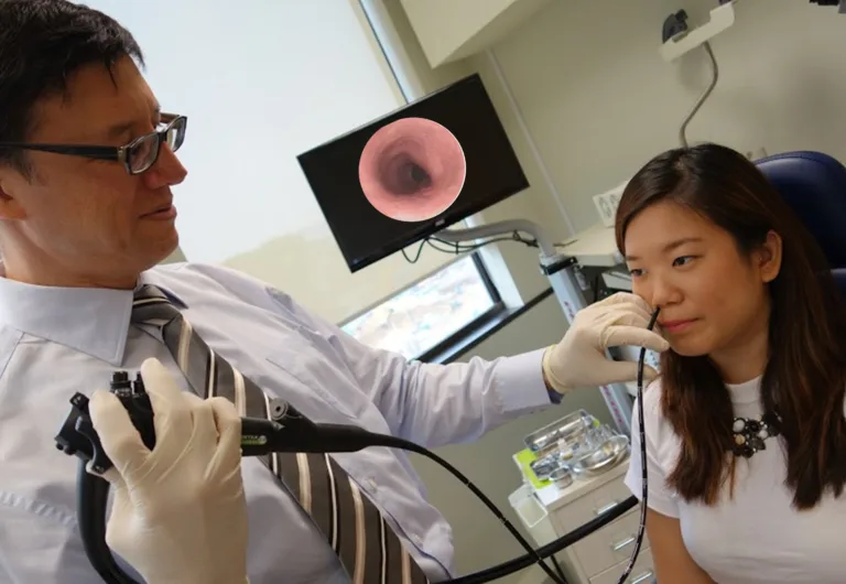
Private Oesophageal Manometry is a test used to assess the function of the oesophagus, the muscular tube that connects the mouth to the stomach. During the test, a thin tube with pressure sensors is inserted through the nose and into the oesophagus. The sensors detect the pressure waves generated by the oesophageal muscles as they contract and relax. This information helps to determine if the oesophageal muscles are functioning normally.

Private Oesophageal Manometry is commonly used to diagnose conditions such as achalasia, a rare disorder that affects the ability of the oesophageal muscles to contract properly. It can also be used to evaluate patients with difficulty swallowing or chest pain.

Reflux Test (24-hour pH and Impedance):

The Reflux Test is a test used to assess the presence and severity of gastroesophageal reflux disease (GERD), a condition in which stomach acid flows back into the oesophagus, causing symptoms such as heartburn and acid regurgitation. The test involves placing a thin tube through the nose and into the oesophagus, which is then attached to a device that measures the pH level (acidic or alkaline) of the oesophagus over a 24-hour period. Additionally, impedance monitoring can be used to measure the movement of liquid or gas in the oesophagus.

This test can help to diagnose GERD and determine the effectiveness of treatment options. It can also be used to evaluate patients with symptoms such as chronic cough, hoarseness, or chest pain.

Private Oesophageal Manometry and Reflux Test Combined:

Private Oesophageal Manometry and Reflux Test Combined is a comprehensive test that combines the two tests described above. It is often used when a patient has symptoms that suggest both oesophageal motility problems and GERD. During the test, a thin tube is inserted through the nose and into the oesophagus to assess the function of the oesophageal muscles (Private Oesophageal Manometry) while also measuring the pH level of the oesophagus over a 24-hour period (Reflux Test).

This test provides a comprehensive evaluation of the function of the oesophagus and can help to diagnose and evaluate treatment options for conditions such as GERD and achalasia.

In conclusion, gastrointestinal physiology tests are important tools that healthcare professionals use to assess the function of the GI tract. Private Oesophageal Manometry, Reflux Test (24-hour pH and Impedance), and Private Oesophageal Manometry and Reflux Test Combined are three commonly used tests that can help diagnose and evaluate treatment options for conditions such as GERD and achalasia. If you are experiencing symptoms related to your GI tract, talk to your healthcare provider to see if a gastrointestinal physiology test is right for you.

Schedule an appointment

What are Gastrointestinal physiology tests ? Read More »

Breath tests for SIBO

What are Breath Tests ?

Procedures

Breath Tests

Breath tests are a non-invasive diagnostic tool used in gastroenterology to diagnose certain digestive conditions. These tests involve measuring the levels of gases in a person’s breath before and after they ingest a small amount of a substance, such as lactose or glucose.

The principle behind breath tests is that when certain substances are digested or fermented by bacteria in the gut, they release gases, such as hydrogen or methane, which can be detected in a person’s breath. By measuring the levels of these gases in the breath, doctors can determine if a person is able to digest or absorb certain substances properly.

Breath tests are commonly used to diagnose lactose intolerance, a condition in which a person’s body is unable to digest lactose, a sugar found in milk and other dairy products. The lactose breath test involves drinking a lactose solution and then measuring the levels of hydrogen and methane in the breath over a period of time.

Other breath tests used in gastroenterology include the glucose breath test, which is used to diagnose small intestinal bacterial overgrowth (SIBO), and the urea breath test, which is used to detect the presence of Helicobacter pylori, a bacteria that can cause stomach ulcers and other digestive problems.

Breath tests are generally safe and non-invasive, and do not involve any radiation or needles. They are also relatively inexpensive compared to other diagnostic tests, and can provide quick results. However, breath tests may not be appropriate for everyone, and their accuracy can be affected by certain medications or medical conditions. It is important to talk to a healthcare provider to determine if a breath test is appropriate and what it can diagnose.

Schedule an appointment

What are Breath Tests ? Read More »

Haemorrhoidal banding

Haemorrhoidal Treatment

Procedures

Haemorrhoidal Treatment

What is Haemorrhoidal banding?

Haemorrhoidal banding is a procedure used to treat symptomatic hemorrhoids. It is a minimally invasive procedure that is performed in a doctor’s office or outpatient setting.

During the procedure, a small rubber band is placed around the base of the hemorrhoid, which cuts off the blood supply to the tissue. The hemorrhoid will then shrink and eventually fall off. This procedure is typically performed under local anesthesia.

Haemorrhoidal banding is considered a safe and effective treatment for symptomatic hemorrhoids, with a low risk of complications. The procedure is typically well tolerated and patients can return to their normal activities soon after the procedure.

It is important to discuss all treatment options with a healthcare provider to determine the best option for each individual patient.

What are Botox injections for anal fissures ?

Botox injections for anal fissures are a non-surgical treatment option for people who have chronic anal fissures that have not responded to other treatments. Botox is the trade name for a type of botulinum toxin, a neurotoxin that works by temporarily paralysing the muscles in the treated area.

In the case of anal fissures, Botox injections are used to relax the internal anal sphincter muscle, which can help to reduce the pain and discomfort associated with the fissure and promote healing. The internal anal sphincter muscle is a ring of smooth muscle that surrounds the anus and controls bowel movements. When this muscle is tense or spasming, it can cause pain and delay healing of an anal fissure.

During the procedure, a small amount of Botox is injected directly into the internal anal sphincter muscle, usually under local anaesthesia. The injection temporarily relaxes the muscle, reducing spasm and improving blood flow to the area, which can help to promote healing of the fissure.

Botox injections for anal fissures have been shown to be effective in relieving pain and promoting healing, with few side effects. However, the effects of Botox are temporary and typically last for a few months. Therefore, repeat injections may be necessary to maintain symptom relief. It is important to note that Botox injections are not appropriate for all patients with anal fissures, and should only be considered after other treatments have been attempted and failed.

Schedule an appointment

Haemorrhoidal Treatment Read More »

Abdominal ultrasound ​

What is a private abdominal ultrasound?

Procedures

Ultrasound

What is an abdominal ultrasound?

A private abdominal ultrasound is an ultrasound examination of the abdomen that is performed by a private clinic or hospital, rather than a publicly funded healthcare facility. Patients typically choose a private abdominal ultrasound for a number of reasons, including:

  • Quicker access to an appointment: Private healthcare facilities can often provide faster access to an ultrasound appointment compared to the public healthcare system, as there may be shorter waiting lists.
  • Increased comfort and privacy: Private clinics and hospitals often offer more comfortable and private facilities compared to the public healthcare system.
  • Increased control over the examination: Patients can choose the type of private abdominal ultrasound they would like to have, and they may be able to request a specific radiologist or sonographer.
  • More detailed reports: Private clinics and hospitals may offer more detailed reports and results compared to the public healthcare system.
  • Increased flexibility: Patients can often choose the time and date of their private abdominal ultrasound appointment, which can be particularly important for those who have busy schedules.

However, it is important to note that private abdominal ultrasounds are typically not covered by healthcare insurance and must be paid for out-of-pocket by the patient. Additionally, private abdominal ultrasounds may not always be necessary, and a referral from a GP or specialist may be required.

What can you see during an abdominal ultrasound?

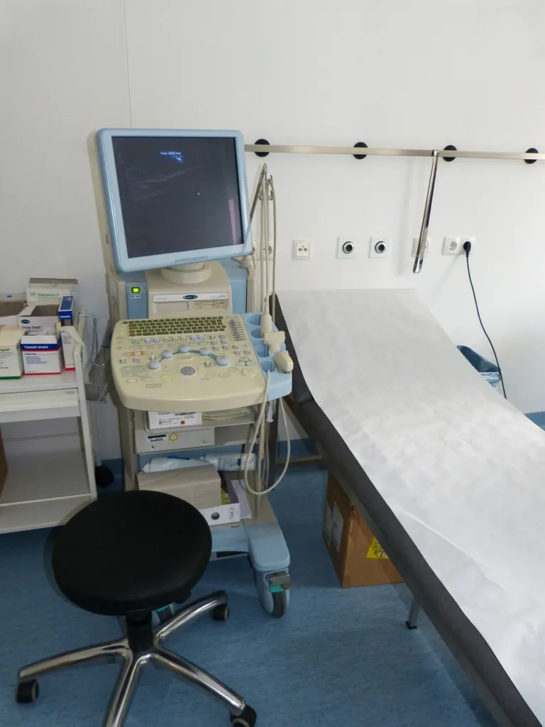
An abdominal ultrasound is a medical imaging test that uses high-frequency sound waves to create images of the organs and tissues within the abdomen. During an abdominal ultrasound, a doctor or a  sonographer (technitian) applies a special gel to the patient’s skin and uses a handheld probe to generate the sound waves and capture the images.

An abdominal ultrasound can provide valuable information about the following organs and structures:

  • Liver: The ultrasound can assess the size, shape, and texture of the liver and detect any abnormalities, such as cysts, tumors, or cirrhosis.
  • Gallbladder: The ultrasound can detect gallstones, evaluate the function of the gallbladder, and identify any inflammation or infection.
  • Pancreas: The ultrasound can evaluate the size and shape of the pancreas and detect any abnormalities, such as cysts or tumors.
  • Spleen: The ultrasound can assess the size and texture of the spleen and detect any abnormalities, such as an enlarged spleen or a mass.
  • Kidneys: The ultrasound can evaluate the size, shape, and texture of the kidneys and detect any abnormalities, such as cysts, tumors, or kidney stones.
  • Abdominal blood vessels: The ultrasound can evaluate the blood flow in the major arteries and veins within the abdomen, such as the aorta and the vena cava.
  • Abdominal fluid collections: The ultrasound can detect the presence of fluid within the abdomen, such as ascites, which can be a sign of liver disease or other underlying conditions.

In addition to these organs, an abdominal ultrasound can also help diagnose other conditions such as hernias, abdominal pain, and digestive tract problems. The images obtained during an abdominal ultrasound can help guide further diagnostic tests or treatments as needed.

What is a pelvic ultrasound ?

A pelvic ultrasound is a non-invasive diagnostic imaging test that uses high-frequency sound waves to produce images of the structures and organs within the pelvic area, such as the uterus, ovaries, bladder, and prostate gland.

In gastroenterology, a pelvic ultrasound may be used to evaluate the lower gastrointestinal tract, including the rectum and anus, and to assess the structures adjacent to it, such as the bladder and prostate in males, and the uterus and ovaries in females.

Some of the common uses of pelvic ultrasound in gastroenterology include:

  1. Evaluating the extent of rectal cancer: Pelvic ultrasound can help determine the size and location of rectal tumors, and can also identify any nearby lymph nodes that may be affected.

  2. Diagnosing pelvic floor disorders: Pelvic ultrasound can help diagnose pelvic floor disorders, such as fecal incontinence or rectal prolapse, by assessing the anatomy and function of the pelvic muscles.

  3. Assessing prostate enlargement: In males, pelvic ultrasound can be used to assess the size of the prostate gland and to identify any abnormalities, such as enlargement due to benign prostatic hyperplasia or the presence of tumors.

  4. Evaluating urinary incontinence: Pelvic ultrasound can be used to evaluate the bladder and urinary tract to determine the cause of urinary incontinence.

Overall, pelvic ultrasound is a useful diagnostic tool in gastroenterology, as it provides detailed images of the pelvic organs and structures, allowing physicians to accurately diagnose and treat a wide range of gastrointestinal conditions.

Schedule an appointment

What is a private abdominal ultrasound? Read More »